
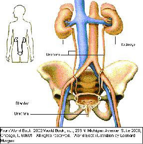
The two ureters are tubes with a length of about 25 cm (10 in)and a diameter of 5 mm (0.2 in). They originate in the renal pelvis (pelvis renalis) of the kidneys (ren), from where they lead through the abdominal wall, cross the blood vessels (vasa sanguinea) of the pelvis and then penetrate the wall of the bladder (vesica urinaria), where they end.
Along this route they transport the urine from the renal pelvis to the bladder. The ureters each comprise three layers. Their insides are lined with a mucous membrane (tunica mucosa). This membrane is followed by a wall of smooth muscle (tunica muscularis). The mucous membrane gives the ureter its elasticity. The layer of muscle promotes transportation of the urine, producing wave-like movements (peristalsis) approx. 1 - 4 times per minute.
A casing of connective tissue (tunica adventitia) forms the outer layer and contains various blood vessels and nerves. It also enables the urethra to move in relation to the organs within its vicinity
Urinary System